![[reticular formation]](./FigVII1.gif)
by Ben Best
Activity of the cerebral cortex is dependent upon both specific
sensory input and nonspecific activating impulses from the brain stem. The
source of these activating impulses is the reticular formation of
the brainstem (ie, medulla, pons and midbrain). The reticular formation
comprises much of the brainstem core, known as the tegmentum. The
reticular formation not only contributes to the activation of the
cortex, but is important for maintaining muscle tone of "antigravity muscles",
assists in regulation of breathing&heartbeat and modulates the sense of pain.
Although the ascending and descending reticular activating systems are well
integrated, the latter tends to be centered in the medulla, whereas the
former is found more in the pons and the midbrain.
![[nuclei of the pons]](./FigVII2.gif)
The reticular formation is actually a loosely arranged network of neurons which are distributed throughout the brainstem wherever there are no specific neural tracts or nuclei. The parvicellular neurons receive inputs from the special senses (which can contribute to arousal. The gigantocellular neurons receive a large portion of inputs from the spine. Pain has a strong influence on reticular activating system activity, particularly in the periaqueductal gray area of the midbrain. Many inputs to the reticular formation arise in higher brain centers, including the cerebellum, hypothalamus, basal ganglia, amygdala and the cerebral cortex (especially the premotor cortex). The gigantocellular neurons give rise to the ascending fibers which travel as the central tegmental tract to the intralaminar nuclei of the thalamus. There is positive feedback between an awake mind and the reticular activating system. The midbrain reticular formation sends projections to the hypothalamus, while paramedian reticular nuclei project mainly to the cerebellum.
Although the content of consciousness is associated with activity of
the cerebral cortex, the reticular activating system is critical for the
existence of the conscious state. Consciousness is lost in both coma and
deep sleep, but these states are very different. Sleep is an active
physiological process for the cerebral cortex, and cerebral oxygen uptake
is comparable to that of the waking state. By contrast, cerebral oxygen
uptake declines in states of coma.
![[midbrain neuromodulator nuclei]](./FigVII3.gif)
Closely associated with the reticular activating system are a number of brain stem nuclei that distribute specific neurotransmitters diffusely to various areas of the brain. Cutting the fibers from the substantia nigra makes cats comatose. Destruction of the locus ceruleus eliminates rapid eye movement (REM) sleep in cats. Destruction of the raphe nuclei results in cats that cannot sleep.
Anatomically, the body of the thalamus is divided by a Y-shaped band of white matter (known as the internal medullary lamina) into three large cell groups: mediodorsal, anterior and lateral. Intralaminar nuclei are found within the lamina itself and a reticular nucleus surrounds the thalamus on the dorsal side.
Functionally, the nuclei of the thalamus can be divided into three
categories: relay nuclei, association nuclei and non-specific nuclei. The
relay nuclei include the geniculate bodies (hearing and vision)
and the ventral nuclei which relay tactile and motor information to the
cerebral cortex. The relay nuclei function to relay sensory and motor
information to the cortex. These nuclei all have reciprocal connections
with the cortex, which undoubtedly gives feedback-control on signals sent.
![[thalamus outputs to the cerebral cortex]](./FigVII5.gif)
![[cortical areas receiving thalamic input]](./FigVII6.gif)
The association nuclei are well-connected with other nuclei in the thalamus, and project largely to the association areas of the frontal and parietal lobes. Three of the four association nuclei are on the dorsal (top) surface of the thalamus: the mediodorsal nucleus, the lateral dorsal nucleus and the lateral posterior nucleus. The fourth association nucleus, the pulvinar, seems to function for general integration of sensory information, with hearing and vision predominating. Evidence indicates that the pulvinar is important for the shifting of attention and suppression of irrelevant inputs.
The non-specific nuclei of the thalamus are the intralaminar nuclei of the reticular nuclei. The intralaminar nuclei are the site of termination for the ascending reticular activating system. The intralaminar neurons project cholinergic fibers diffusely to the cerebrum providing generalized activation. They also project to the striatum (caudate and putamen) of the basal ganglia.
The thalamic reticular nucleus (complex) is really only a thin sheet
of inhibitor neurons which are believed to function to "gate" signals to the
cerebrum from the thalamus. Most thalamic neurons have few, if any, collaterals,
but the reticular neurons have extensive collaterals. Since projections to
the cortex from the thalamus must pass through the reticular nucleus complex,
it is believed that this complex acts as a "gateway". The reticular complex
may control which sensory inputs are the subject of attention of the
cerebral cortex.
![[relation of thalamic reticular complex to cortex and
thalamus]](./FigVII7.gif)
The membranes of the thalamic neurons sending projections to the cerebrum have specialized properties which enable them to fire in synchronized bursts or with desynchronized single-spike firing. The former corresponds to the EEG desynchronization seen in REM sleep or in the alert, awakened state. Projections from the reticular complex may determine the mode of firing of thalamic neurons.
The cerebellum is functionally and anatomically divided into three
parts: the archicerebellum (consisting of the physically separated
flocculonodular lobe), the paleocerebellum (anterior lobe) and
the neocerebellum (posterior lobe). THe archicerebellum is the oldest
part from an evolutionary point of view, and it is part of the vestibular
system — concerned with balance and equilibrium.
|
The paleocerebellum is most concerned with regulation of muscle tone. It receives inputs from muscle stretch receptors via a distinctive structure in the medulla known as the inferior olive (which looks like a crumpled-up bag). The inferior olive also receives inputs from a number of midbrain nuclei such as the superior colliculus and the red nucleus. The inferior olive sends outputs to the cerebellum through the inferior cerebellar peduncle.
The neocerebellum is the largest&newest part of the cerebellum —
and it receives inputs from the cerebral cortex via the pontine nuclei in
the base of the pons. Axons from the pontine nuclei enter the cerebellum
through the middle cerebellar peduncles. The major output tract of
the cerebellum is the superior cerebellar peduncle, which primarily
sends signals to the motor cortex and the supplementary motor area.
![[cerebellar cortex neuron layers]](./FigVII10.gif)
The cerebellar cortex consists of three layers with five types of
neurons: stellate, basket, Purkinje, Golgi and
granule cells. The outermost molecular layer is composed
primarily of granule cell axons and Purkinje cell dendrites. The middle
Purkinje cell layer contains the large Purkinje cells arranged
side-by-side in a single layer. The innermost granular layer is
primarily composed of approximately 100 billion small granule cells —
more cells than is found in the entire cortex.
![[cerebellum pathways in the cerebellum]](./FigVII11.gif)
The only output of the cerebellum is from the Purkinje cells, which
in turn are controlled by two types of input: mossy fibers and
climbing fibers. Mossy fibers end on granule cell dendrites, and the
granule cells send large numbers of axons to the Purkinje cells. A very large
number of active mossy fibers are necessary to cause a Purkinje cell to fire.
By contrast, there is one climbing fiber for about ten Purkinje cells, and
every single action potential in a climbing fiber will cause all ten (or so)
Purkinje cells to fire. Whereas mossy fibers can come from many sources, every
climbing fiber comes from a single cell in the inferior olive. Mossy fibers
give graded response, whereas climbing fibers provide an all-or-nothing
action. There is evidence to indicate that climbing fibers provide error
signals in the execution of movement.
![[cerebellum pathways to the cortex]](./FigVII12.gif)
The cerebellum and its associated brain stem nuclei function to compare the intention of motor movement with actual performance, and to make corrections when there is a mismatch. The cerebellum is particularly important for controlling the balance between antagonistic muscle groups during rapid changes in body position, including such activities as running or playing the piano. The cerebellum has extraordinary computational capabilities with respect to its ability to calculate the anticipated positions of rapidly moving body parts. And the cerebellum can learn from its mistakes when movements do not occur as intended.
The basal ganglia are a collection of forebrain nuclei at the base
of the cerebral hemispheres (early anatomists referred to the nuclei as
"ganglia"). The caudate nucleus is an elongated extension of the
putamen. Closely associated with these two structures is the
nucleus accumbens. All three of these nuclei are collectively
referred to as the striatum.
![[basal ganglia detailed structure]](./FigVII14.gif)
![[basal ganglia functional interconnections]](./FigVII15.gif)
All inputs to the basal ganglia come from the cerebral cortex and enter at the striatum. Spiny neurons with axons that form radial fibers (resulting in a striated, or striped, appearance) constitute 96& 37; of striatal neurons. The striatum surrounds the globus pallidus ("pale sphere") consisting of an internal and external segment. The other nuclei of the basal ganglia are the subthalamus and the substantia nigra (although anatomically the latter lies in the midbrain).
Each of the nuclei of the striatum serves as the input point for signal circuits that originate in the cerebral cortex, pass through other basal ganglia and then return to the cerebral cortex via the thalamus. The putamen receives input for a "sensorimotor loop". The caudate receives input for an "association loop". And the nucleus accumbens receives input for a "limbic loop". The looplike character of these circuits undoubtedly accounts for the fact that oscillation is a common feature of basal ganglia damage.
The putamen loop (sensorimotor loop) from the premotor and somatosensory areas of the cortex is deemed responsible for facilitating subconscious execution of learned movements. Cutting paper with scissors, throwing a baseball, hammering a nail or even writing are subserved by this loop. PET scans show putamen activity in the learning of complex motor sequences. The circuit evidently facilitates the execution of motor programs stored in the cortex, because monkeys with lesions in the globus pallidus retain their ability to perform motor tasks — although they do so more slowly. Lesions to the globus pallidus can also lead to spontaneous writhing movements, whereas lesions to the subthalamus often results in sudden violent flailing of a limb.
The caudate loop (association loop) begins with inputs from the association areas of the cerebral cortex, rather than the sensory or motor areas. The caudate loop is regarded as being important for cognitive control&planning of movement sequences and movements executed in parallel (synchronously — "walking and chewing gum at the same time"). Running to a tree and climbing it to escape from a wild beast has been given as an example of the caudate loop in action. Route-finding is associated with caudate activity in PET scans. Because the caudate circuit is implicit in the timing&scaling of movements, writing in small or large letters on a blackboard is also cited as a function mediated by the caudate.
Both the sensorimotor and association loops return to the cerebrum via the ventrolateral nucleus of the thalamus (the same region of the thalamus relaying output from the cerebellum), as well as the ventroanterior nucleus of the thalamus. By contrast, the limbic loop returns signals to the cerebrum via the mediodorsal thalamic nucleus. Receiving inputs from the amygdala and the cingulate gyrus, the limbic loop may function in the motor expression of emotions (like smiling or assuming an aggressive posture) or in the motivational aspect of alteration of movements.
The basal ganglia are thought to be important when movements are learned by repetition rather than by insight. Many striatum neurons are most active when responding to a stimulus linked to memories of significant events, which has led to the suggestion that the basal ganglia link motivation to the execution of movements. The striatum is associated with the initiation and termination of action sequences, habit learning and reward-associated (including punishment-avoiding) learning.
Patients with carbon monoxide damage to the globus pallidus and putamen show a loss of initiative and a tendency to engage in repetitive activities, even when there is no evident loss of intellectual capacity. These patients often use ritualized phrases in association with certain behaviors, and frequently count to themselves and/or snap their fingers. One patient could spend 15 minutes flipping a light-switch on and off, whereas another would not utter a sentence until she was certain that the number of words was a multiple of 3. I speculate that the high carbon monoxide content of tobacco smoke may contribute to the smoking habit by basal ganglia damage leading to ritualized puffing and lighting of cigarettes.
The striatum receives tonic inhibition from dopamine-containing axons originating in the melanin-containing neurons of the substantia nigra. 80 % of the dopamine in the brain is found in the basal ganglia (which account for half-a-percent of the total brain weight). Degeneration of dopaminergic neurons to the extent that dopamine content of the striatum is reduced 80 %-90 % leads to Parkinson's Disease. A Parkinson patient demonstrates tremor, rigidity and intense willful effort required for even routine activities like opening a door. By contrast, patients with Huntington's Chorea suffer from hyperactivity of dopamine in the striatum, resulting in choreiform (dancelike) movements.
The body of an animal can be divided into somatic and visceral portions.
The somatic portion interacts with the external environment through the
skeletomuscular system under the control of the cerebral cortex. The internal
environment (inside the body) is regulated by the autonomic nervous system
under supervision of the hypothalamus and brain stem nuclei. Specifically,
the Autonomic Nervous System (ANS) supervises heartbeat, digestion, defecation
reflexes, blood vessel constriction, etc. The word "autonomic" implies that the
ANS is independent of the cerebral cortex and not subject to conscious control.
This is largely true, but people have trained themselves to gain conscious
control of their heart rates, for example.
![[autonomic nervous system]](./FigVII16.gif)
The ANS is divided into sympathetic and parasympathetic divisions. The parasympathetic division supervises the truly visceral functions, through ganglia located directly in organs such as the kidney, liver, gut, etc. Just as the central nervous system controls voluntary muscles by the release of acetylcholine, the parasympathetic nervous system releases acetylcholine into digestive secretory glands, heart muscle and smooth muscle (such as the muscles controlling the slow contractions of the intestine). Smooth muscle differs from skeletal muscle insofar as the response to stimulation can last for minutes, hours or days — as opposed to a fraction of a second. Smooth muscles contract 100 to 1,000 times more slowly than skeletal muscles. Skeletal muscle receptors are called nicotinic because nicotine can mimic acetylcholine, whereas parasympathetic end-organs are called muscarinic because the toadstool poison muscarine (not nicotine) mimics acetylcholine. Parasympathetic nerves arise from the top and bottom of the spinal cord, as opposed to sympathetic nerves, which arise from the central portions of the spinal cord.
While the parasympatheic division of the ANS handles vegetative functions, the sympathetic division is more concerned with mobilization of body resources to handle stress. The sympathetic division most often functions by mass discharge with simultaneous effects upon the entire body, rather than by isolated target organs. The sympathetic ganglia are located close to the spine rather than in target organs. And sympathetic nerves release norepinephrine (noradrenalin) on target organs, rather than acetylcholine. The adrenal medulla (interior portion of the adrenal gland) is actually a very specialized sympathetic ganglion which releases epinephrine (adrenalin) into the blood stream. Both epinephrine and norepinephrine increase blood glucose, increase the rate&force of heart contraction and increase metabolic rate. Norepinephrine constricts all arterioles, whereas epinephrine only dilates skeletal muscle blood vessels (both effects increase blood volume in muscles). Epinephrine dilates the bronchi of the lungs.
Sympathetic response has often been described as preparation for
"fight or flight", associated with anger and fear. Insofar as male
ejaculation is also under sympathetic control, another "f-word" could be
added to the list. Epinephrine, however, is more associated with fear than
is norepinephrine. Arterioles can be constricted by the sympathetic
nervous system, whereas about the only blood vessels innervated by the
parasympathetic nervous system are the ones in the genitals associated
with the blood engorgement of sexual arousal. (Observe that an erection can
be produced by genital stimulation or by thinking sexual thoughts, but not
by a direct act of will, like extending an arm). Blushing and the
paleness of fright are examples of sympathetic emotional expression through
action on blood vessels. Sympathetic activity can make hairs stand up, and
this is of more significance in animals than in humans.
![[functional nuclei in the hypothalamus]](./FigVII17.gif)
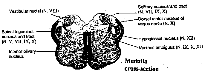
Bowel emptying is largely a matter of local parasympathetic reflex activity. Swallowing, coughing, sneezing, gagging and vomiting are all reflex responses integrated in the medulla. The solitary nucleus of the medulla is the major coordinating center for autonomic activity in the brain stem. Pressure sensors in the aortic arch, for example, send sensory signals to the solitary nucleus which can result in reflex slowing of heart rate. But the highest center for the autonomic nervous system is the hypothalamus.
The hypothalamus is not only the center of the autonomic nervous
system, it is the center of the body's hormonal system and is important
for the expression of emotion. The hypothalamus does not exercise control
in the same direct way as the CNS affects the somatic system, but regulates
more through bias and modulation. To regulate effectively, the hypothalamus
must receive inputs from many diverse brain areas, as shown in the illustration.
![[hypothalamic nuclei in detail]](./FigVII19.gif)
The hypothalamus functions to regulate body temperature, hunger, thirst, osmotic pressure, sex drive, etc. The suprachiasmatic nucleus receives fibers emerging from the optic nerve and seems to function in the regulation of circadian rhythms ("day-night cycles"). Signals from the suprachiasmatic nucleus to the pineal gland results in the production of melatonin from serotonin. Stimulation of parts of the median forebrain bundle in male monkeys can result in penile erection and emotional display. Stimulation of the periventricular nucleus (or of the periaqueductal gray area, with which this nucleus is continuous) leads to fear, escape or punishment reactions. (The periaqueductal gray, also known as central gray, is a heterogenous collection of gray matter surrounding the cerebral aqueduct in the midbrain. It also has analgesic and reproductive functions, as well as others). (The periventricular nucleus should not be confused with the paraventricular nucleus.) The mammillary bodies in the posterior portion of the hypothalamus receive inputs mainly from the hippocampus via the fornix, and send outputs to the reticular formation and to the cingulate cortex via the mediodorsal thalamic nucleus.
Stimulation of the ventromedial nucleus causes cessation of eating, docile behavior and aversive reactions, whereas destruction of this nucleus results not only in gross overeating, but in savage destructiveness. Control of eating, however, cannot be localized to a single hypothalamic nucleus. Bilateral lesions to the lateral hypothalamic area (LHA) cause a rat to stop eating and drinking. Moreover, the rats are lethargic and have a limited tolerance for stress. Studying the isolated function of the LHA is made difficult by the fact that LHA neurons are diffusely arranged within an area traversed by the median forebrain bundle (MFB). The MFB is the main conduction pathway linking the brainstem (reticular formation, locus ceruleus, dorsal raphe and ventral tegmental area), hypothalamus and basal forebrain (amygdala and striatum). A hungry rat will press a lever at a rate of once per second to electrically stimulate the LHA, but a meal or glucose infusion inhibits this action. Dopamine released into the nucleus accumbens and prefrontal motor cortex is the reward for feeding. The paraventricular nucleus causes hunger for carbohydrate prior to feeding and, as feeding progresses, shifts the preference from carbohydrate to fat. Cholecystokinin (CCK) promotes carbohydrate satiety and dopamine inhibits fat consumption.
Of the endocrine organs, only the adrenal medulla is controlled by
direct innervation from the autonomic nervous system. The pituitary gland is
the "master gland" regulating the body's endocrine system by means of "releasing
factors". The anterior pituitary is itself controlled by control hormones
released from within the hypothalamus, whereas the posterior pituitary is
controlled by nerve signals from the hypothalamus. One posterior pituitary
hormone, oxytocin, helps deliver milk from the breast glands to the
nipples. A sucking stimulus to the nipples — or even the sight or sound of
a mother's infant — can result in a pulse of oxytocin to the mammary gland
within 13 seconds. Sex hormones and thyroid hormones pass the blood-brain
barrier easily. Sex hormone receptors have been found on hypothalamic cells
as well as on cells elsewhere in the brain, particularly certain limbic
structures.
![[functional actions of hypothalamus and pituitary]](./FigVII20.gif)
Psychological stress can depress the immune system, and this effect is mediated through the hypothalamus. High anxiety and stress increases AdrenoCorticoTropic Hormone (ACTH) and corticosteroids which, in turn, reduces phagocytic, B and T lymphocyte proliferation in response to an antigen. Evidence for this action is supported by animal experiments which demonstrate reduced lymphocytic responses to antigen in rats subjected to tail shocks or restraining apparatus. Moreover, immune mediators like cytokines are released from brain cells and affect pituitary hormone release.
|
Chapter 3 of this monograph described the gross function of the hippocampus in detail, but a few additional anatomical and functional notes should be made.
|
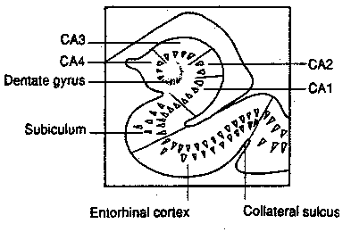
The hippocampus is also known as Ammon's horn (Cornu
Ammonis) because of its resemblance to an Egyptian deity having the head of
a ram. The hippocampus is divided into four "CA" zones, of which CA1 and CA3
are the largest. Although the hippocampus is a continuation of the cerebral
cortex, it contains three layers rather than six: molecular layer,
granular layer and polymorphic layer. "Hippocampus" is Greek
for "seahorse", and the "nose" of the seahorse is a structure called the
fimbria, which is the beginning of the fornix. The fornix is
a bundle of fibers connecting the hippocampus with the septal nuclei and with
the mammillary bodies of the hypothalamus.
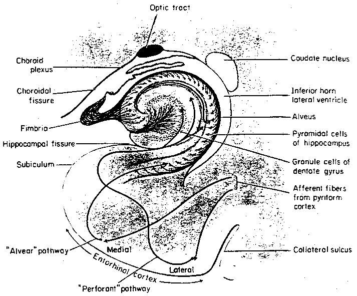
![[the fornix and its connections]](./FigVII23.gif)
In the 1930s a neurologist named Papez proposed a "circuit" of emotion that included the thalamus, mammillary bodies, cingulate cortex and hippocampus, but did not include the amygdala. Although rabies particularly affects the hippocampus, the hippocampus is currently regarded as an organ of learning rather than an organ of emotion. The fornix contains fibers that connect the hippocampus with the septal nuclei ("pleasure centers"), but most of the output of hippocampal processing apparently goes to the subiculum, which is the superior portion of the parahippocampal gyrus of the temporal lobe. Most of the fibers of the fornix travel from the subiculum to the mammillary bodies. The subiculum also sends extensive projections to the association areas of the frontal and parietal lobes. Lesions in the mammillary bodies and the medial dorsal nucleus of the thalamus are found in Korsakoff's syndrome, which involves severe memory impairment without a clouding of consciousness. As in the case of patients with bilateral hippocampal damage, Korsakoff patients have difficulty with learning and recent memory, rather than with long-term memories formed before the onset of the condition.
![[gross outline of the limbic system]](./FigVII24.gif)
![[detailed limbic system structures]](./FigVII25.gif)
|
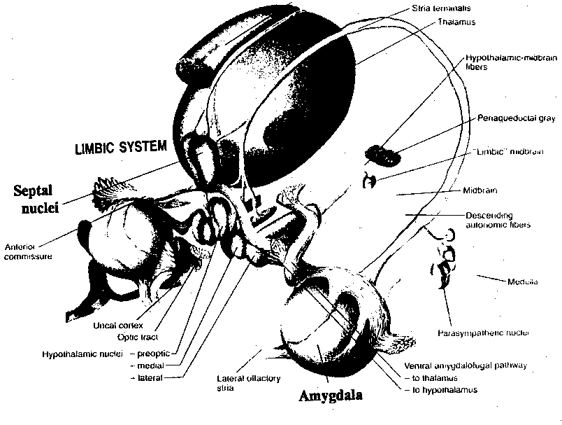
"Limbic system" is a term that has come to mean those parts of the brain most concerned with emotion. That definition should include the hypothalamus and probably exclude the hippocampus, but there is no universally agreed-upon list of included structures. This review will focus on the septal area and the amygdala.
The septal area consists of the septal nuclei merged with the cortex directly in front of the anterior commissure. Its major connections are through the fornix, but it also receives inputs from the amygdala and has reciprocal connections with the hypothalamus. The septal area is an important pleasure area of the brain, and laboratory animals eagerly press levers that send electrical impulses to this area. Electrical stimulation of the septal area has an inhibitory effect on the autonomic nervous system, including cardiac deceleration. Septal lesions produce rage reactions in many species, but these seem to be defensive rather than aggressive because the animals try to avoid unfamiliar situations and conflict. Other species become less aggressive following septal lesions.
The entorhinal cortical area, the most posterior part of the
pyriform (primary olfactory cortex) lobe, corresponds to area 28 of
Brodmann, and constitutes a major portion of the anterior parahippocampal
gyrus in humans. The amygdaloid nuclear complex is caudally (posteriorly)
continuous with the uncus ("hooked" portion) of the parahippocampal gyrus.
The amygdala ("almond") is the central structure of the limbic system.
![[main parts of the amygdala]](./FigVII27.gif)
![[main connections of the amygdala]](./FigVII28.gif)
The
amygdala
is divided into a centromedial[Ce-M] (or
centrocortical[Ce-Co]), part concerned with autonomic functions and a
basolateral[BL] part concerned with conscious processing. The
centromedial portion receives inputs directly from the hypothalamus[H} and
septal area[S]. Both portions of the amygdala send outputs to the septal area
and the hypothalamus via a long band of fibers (arching around the striatum
and internal capsule) known as the stria terminalis. A collection of
neurons known as the basal nucleus of the stria terminalis[BST] is
closely associated with the centromedial nucleus as a dumbbell-shaped
structure known as the extended amygdala.
![[limbic brain stem connections]](./FigVII29.gif)
Stimulation of the extended amygdala may result in smacking, salivation, licking and chewing movements. There may also be emptying of the bladder and rectum. Stimulation of the corticomedial amygdala increases food intake, whereas stimulation of the basolateral amygdala reduces feeding behavior.
Stimulation of the basolateral nuclear group results in arousal
and attention. Strong stimulation of the same group can produce powerful
fear or rage. Destruction of the amygdala of violent criminals has reduced
their aggressiveness. The amygdala seems to be important for learning to
associate objects with reward or punishment. The amygdala is also a key
structure for fear learning. Fear conditioning has been demonstrated to have
many effects on hypothalamic and brain stem centers mediated by the central
nucleus of the amygdala.
![[targets and influences of the amygdala]](./FigVII30.gif)
The amygdala is among the brain areas with the highest density of receptors for sex hormones. Stimulation of the corticomedial amygdala may cause ovulation in the female, but cutting the stria terminalis abolishes this effect. The range of emotions associated with the amygdala is really quite wide.
![[thalamus nuclei]](./FigVII4.gif)
![[cerebellum]](./FigVII8.gif)
![[basal ganglia location in the brain]](./FigVII13.gif)
![[pathways in the hypothalamus]](./FigVII18.gif)
![[GO TO BEN BEST'S HOME PAGE]](../../homeback.gif) HOME PAGE
HOME PAGE